Home
Uncategories
Lentigo Maligna Melanoma Histology - Histologic Criteria For Diagnosing Primary Cutaneous Malignant Melanoma Modern Pathology : Lentigo maligna is a precursor to lentigo maligna melanoma, a potentially serious form of skin cancer.
Lentigo Maligna Melanoma Histology - Histologic Criteria For Diagnosing Primary Cutaneous Malignant Melanoma Modern Pathology : Lentigo maligna is a precursor to lentigo maligna melanoma, a potentially serious form of skin cancer.
Lentigo Maligna Melanoma Histology - Histologic Criteria For Diagnosing Primary Cutaneous Malignant Melanoma Modern Pathology : Lentigo maligna is a precursor to lentigo maligna melanoma, a potentially serious form of skin cancer.. Lentigo maligna melanoma is a melanoma that has evolved from a lentigo maligna,:695 as seen as a lentigo maligna with melanoma cells invading below the boundaries of the epidermis. Biopsy and histology may be used to differentiate the various types of lentigines. It appears as a brown or black mottled, irregular, lesion with increased numbers of scattered atypical melanocytes in the epidermis. Irregular patch about 10mm square after scrape biopsy which concluded suspicious of early malignant melanoma. Lentigo maligna grows slowly and is usually harmless, but lentigo maligna melanoma can spread aggressively.
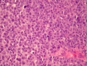
Lentigo maligna melanoma (including lentigo maligna). Colour before scrape biopsy was light brown. Lentigo maligna grows slowly and is usually harmless, but lentigo maligna melanoma can spread aggressively. Biopsy and histology may be used to differentiate the various types of lentigines. A melanoma of the skin characterized by single cell infiltration of the papillary dermis by atypical melanocytes, in a background of lentigo maligna changes.
Melanoma Pathology Dermnet Nz from dermnetnz.org Lentigo simplex may also be confused with a paucicellular lentiginous junctional melanocytic nevus, including nevus spilus, in which a nest is seen only rarely and may be missed unless several levels are examined. Biopsy and histology may be used to differentiate the various types of lentigines. Melanoma lentigo maligna lentigo maligna melanoma treatment dermoscopy reflectance confocal microscopy diagnosis treatment staged excision. Lentigo maligna is a precursor to lentigo maligna melanoma, a potentially serious form of skin cancer. They are usually found on chronically sun damaged skin such as the face and the forearms of the elderly. Lentigo maligna melanoma should be completely removed surgically. Conversion to a lentigo maligna melanoma can take investigations5. J am acad dermatol 1995;
It is rare for a lentigo simplex to be mistaken for melanoma in situ, but it may happen.
Lentigo maligna and lentigo maligna melanoma. Ssmm is actually diagnosed by a distinctive pattern displayed by tumour cells within the epidermis. Lentigo maligna grows slowly and is usually harmless, but lentigo maligna melanoma can spread aggressively. This cancer typically starts on the surface of the skin on parts of the body that. Lentigo maligna melanoma represents 4% to 10% of melanomas; A website about lentigo maligna melanoma. Biopsy and histology may be used to differentiate the various types of lentigines. Lentigo maligna is a precursor to lentigo maligna melanoma, a potentially serious form of skin cancer. May arise secondary to lentigo maligna. It develops from lentigo maligna, also known as hutchinson's melanotic freckle, which is a slow growing type of melanoma. The tumors are often larger than 3 cm, flat, and tan, with marked notching of the borders; Lentigo maligna (a subtype of melanoma in situ), by definition, does not infiltrate into dermis but lentigo maligna melanoma has at least single cell infiltration into papillary dermis. Lentigo maligna is not the same as lentigo maligna melanoma, as detailed below.
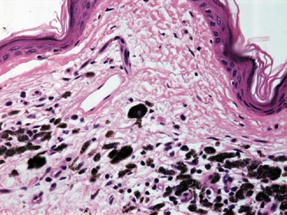
Working in a histology lab means that i get to see a lot of what our body looks like under the microscope. Tannous zs, lerner lh, duncan lm, et al. Although these melanomas spread peripherally before invading deeper, there is often a delay in diagnosis because. It appears as a brown or black mottled, irregular, lesion with increased numbers of scattered atypical melanocytes in the epidermis. However, lesions on the face can be difficult to interpret, for more information please refer to the related chapter on melanoma:
Lentigo Maligna Melanoma Sciencedirect from ars.els-cdn.com Although regression is a prognostic factor, the histologic type is more important for histology coding purposes. Lentigo maligna is also known as hutchinson melanotic freckle. Lentigo maligna grows slowly and is usually harmless, but lentigo maligna melanoma can spread aggressively. Melanocytes are the cells responsible for making melanin, the pigment that determines the color of the skin. Although these melanomas spread peripherally before invading deeper, there is often a delay in diagnosis because. J am acad dermatol 1995; They are usually found on chronically sun damaged skin such as the face and the forearms of the elderly. Lentigo maligna melanoma (including lentigo maligna).
It develops from lentigo maligna, also known as hutchinson's melanotic freckle, which is a slow growing type of melanoma.
Photographic documentation of a single case. May arise secondary to lentigo maligna. This cancer typically starts on the surface of the skin on parts of the body that. The visual symptoms of lentigo maligna melanoma are very similar to those of lentigo maligna. Lentigo maligna (lm) and lentigo maligna melanoma (lmm) are types of skin cancer. It is characterized by a highly irregular border, heterogeneous coloration, and—because of its long radial growth phase—a large diameter. The doctor should remove a long ellipse of skin, take biopsies from multiple sites or carefully shave a representative area for histology. Conversion to a lentigo maligna melanoma can take investigations5. Lentigo maligna melanoma is a melanoma that has evolved from a lentigo maligna,:695 as seen as a lentigo maligna with melanoma cells invading below the boundaries of the epidermis. Is a specific histologic type of in situ melanoma. Lentigo maligna melanoma represents 4% to 10% of melanomas; A melanoma of the skin characterized by single cell infiltration of the papillary dermis by atypical melanocytes, in a background of lentigo maligna changes. Lentigo maligna melanoma (lmm) = lentiginous malignant melanoma with sun damage.
Biopsy and histology may be used to differentiate the various types of lentigines. The tumors are often larger than 3 cm, flat, and tan, with marked notching of the borders; Lentigo maligna is also known as hutchinson melanotic freckle. Tannous zs, lerner lh, duncan lm, et al. Lentigo maligna and lentigo maligna melanoma.
Histologic Criteria For Diagnosing Primary Cutaneous Malignant Melanoma Modern Pathology from media.springernature.com It is rare for a lentigo simplex to be mistaken for melanoma in situ, but it may happen. Histologic similarities between lentigo maligna and dysplastic nevus: Photographic documentation of a single case. They begin when the melanocytes in the skin grow out of control and form tumors. This cancer typically starts on the surface of the skin on parts of the body that. Dence of cutaneous melanoma in the united states by. Colour before scrape biopsy was light brown. Although regression is a prognostic factor, the histologic type is more important for histology coding purposes.
Lentigo maligna is a precursor to lentigo maligna melanoma, a potentially serious form of skin cancer.
Biopsy and histology may be used to differentiate the various types of lentigines. It is rare for a lentigo simplex to be mistaken for melanoma in situ, but it may happen. Lentigo maligna and lentigo maligna melanoma. Lentigo maligna is also known as hutchinson melanotic freckle. It develops from lentigo maligna, also known as hutchinson's melanotic freckle, which is a slow growing type of melanoma. A melanoma of the skin characterized by single cell infiltration of the papillary dermis by atypical melanocytes, in a background of lentigo maligna changes. It aims to provide a sequence of photographs that demonstrate the appearance of the first lesion, and then its progression — including the diagnostic biopsy and the subsequent plastic surgery. They are usually found on chronically sun damaged skin such as the face and the forearms of the elderly. The tumors are often larger than 3 cm, flat, and tan, with marked notching of the borders; Lentigo maligna melanoma is a relatively uncommon form of skin cancer that tends to affect older individuals. J am acad dermatol 1995; Lentigo maligna melanoma (including lentigo maligna). Ssmm is actually diagnosed by a distinctive pattern displayed by tumour cells within the epidermis.

0 Comments:
Posting Komentar