Home
Uncategories
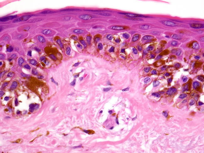
Lentigo Histology : Lentigo Maligna Melanoma Overview Etiology And Pathophysiology Epidemiology : Histologic findings may include hyperplasia of the histologic findings may include hyperplasia of the epidermis and increased pigmentation of the basal layer.
Lentigo Histology : Lentigo Maligna Melanoma Overview Etiology And Pathophysiology Epidemiology : Histologic findings may include hyperplasia of the histologic findings may include hyperplasia of the epidermis and increased pigmentation of the basal layer.
Lentigo Histology : Lentigo Maligna Melanoma Overview Etiology And Pathophysiology Epidemiology : Histologic findings may include hyperplasia of the histologic findings may include hyperplasia of the epidermis and increased pigmentation of the basal layer.. Lentigo and junctional nevus can look like a freckle. In contrast, freckles darken after sun exposure and are present from early childhood. Harriet cheng bhb, mbchb, dermatology department a lentiginous naevus (also called naevoid lentigo or 'jentigo') has few junctional nests and lentiginous melanocytic hyperplasia at the periphery. Histologic findings may include hyperplasia of the histologic findings may include hyperplasia of the epidermis and increased pigmentation of the basal layer. Lentigo simplex is acquired in childhood, but the lentigines are not confined to.
Pronounced pigmention of the tips of elongated rete. Reflects the particular epidermal architecture marked by. Tanning bed lentigo appears after exposure to an indoor tanning bed. Of ink spot lentigo showing lentiginous hyperplasia of. The epidermis, marked hyperpigmentation of the basal.
Lentigo Maligna Wikipedia from upload.wikimedia.org Of ink spot lentigo showing lentiginous hyperplasia of. Lentigo simplex is acquired in childhood, but the lentigines are not confined to. The epidermis, marked hyperpigmentation of the basal. Puva lentigo starts after psoralen and ultraviolet a (puva) therapy, which is used to treat conditions like eczema and psoriasis. Large numbers of lentigines are very common, especially in patients with red hair. Lentigines (plural of lentigo) are flat brown lesions which do not darken following sun exposure (thus differentiating them from ephelides, or true freckles). Puva (psoralen + ultraviolet a) treatment for psoriasis cause numerous solar lentigines with atypia; Lentigo simplex is a common benign melanocytic lesion.
Lentigo and junctional nevus can look like a freckle.
A variable number of melanocytes are. Similar findings after severe radiation exposure. Lentigines (plural of lentigo) are flat brown lesions which do not darken following sun exposure (thus differentiating them from ephelides, or true freckles). Puva (psoralen + ultraviolet a) treatment for psoriasis cause numerous solar lentigines with atypia; Histologic findings may include hyperplasia of the histologic findings may include hyperplasia of the epidermis and increased pigmentation of the basal layer. A/prof patrick emanuel, dermatopathologist, auckland, new zealand; Lentigo maligna melanoma has no better prognosis than other types of melanoma. Puva lentigo starts after psoralen and ultraviolet a (puva) therapy, which is used to treat conditions like eczema and psoriasis. Multiple large solar lentigos on upper back and shoulders suggest prior severe sunburn, a risk factor for melanoma (dermatology 2007;214:25). They can be distinguished from freckles by their darker colour, wider distribution and the fact that they do not disappear in winter months. Of ink spot lentigo showing lentiginous hyperplasia of. Lentigo and junctional nevus can look like a freckle. Microscopy of ink spot lentigo is pathognomonic and.
Lentigines (plural of lentigo) are flat brown lesions which do not darken following sun exposure (thus differentiating them from ephelides, or true freckles). Little attention is paid to this insidious lesion which can potentially become an invasive lentigo maligna koh hk, michalik e, sober aj: It is a harmless (benign) hyperplasia of melanocytes which is linear in its spread. A variable number of melanocytes are. Lentigo maligna melanoma has no better prognosis than other types of melanoma.
Webpathology Com A Collection Of Surgical Pathology Images from www.webpathology.com Of ink spot lentigo showing lentiginous hyperplasia of. Reflects the particular epidermal architecture marked by. Lentigo simplex is a common benign melanocytic lesion. Pronounced pigmention of the tips of elongated rete. Multiple large solar lentigos on upper back and shoulders suggest prior severe sunburn, a risk factor for melanoma (dermatology 2007;214:25). It is also known as simple lentigo. Little attention is paid to this insidious lesion which can potentially become an invasive lentigo maligna koh hk, michalik e, sober aj: Lentigo and junctional nevus can look like a freckle.
A/prof patrick emanuel, dermatopathologist, auckland, new zealand;
Histologic findings may include hyperplasia of the histologic findings may include hyperplasia of the epidermis and increased pigmentation of the basal layer. Microscopy of ink spot lentigo is pathognomonic and. It is also known as simple lentigo. Reflects the particular epidermal architecture marked by. The epidermis, marked hyperpigmentation of the basal. Radiation lentigo happens in areas of the skin that have been exposed to radiation — for example. Similar findings after severe radiation exposure. Lentigines (plural of lentigo) are flat brown lesions which do not darken following sun exposure (thus differentiating them from ephelides, or true freckles). Lentigo simplex is acquired in childhood, but the lentigines are not confined to. Large numbers of lentigines are very common, especially in patients with red hair. They can be distinguished from freckles by their darker colour, wider distribution and the fact that they do not disappear in winter months. May be precursor to seborrheic keratosis. A variable number of melanocytes are.
Actinic lentigo does not darken with sun exposure and is acquired later in life. A variable number of melanocytes are. Lentigo simplex is a common benign melanocytic lesion. Lentigo maligna melanoma has no better prognosis than other types of melanoma. This single lesion of the upper back contains both lentigo maligna, an in situ histology, and superficial spreading melanoma, an invasive histology.
Daub Discolouration Pigmentation Solar Lentigo from www.heighpubs.org Microscopy of ink spot lentigo is pathognomonic and. Puva lentigo starts after psoralen and ultraviolet a (puva) therapy, which is used to treat conditions like eczema and psoriasis. The epidermis, marked hyperpigmentation of the basal. Radiation lentigo happens in areas of the skin that have been exposed to radiation — for example. Tanning bed lentigo appears after exposure to an indoor tanning bed. Reflects the particular epidermal architecture marked by. Histologic findings may include hyperplasia of the histologic findings may include hyperplasia of the epidermis and increased pigmentation of the basal layer. A variable number of melanocytes are.
Similar findings after severe radiation exposure.
Radiation lentigo happens in areas of the skin that have been exposed to radiation — for example. Multiple large solar lentigos on upper back and shoulders suggest prior severe sunburn, a risk factor for melanoma (dermatology 2007;214:25). Puva lentigo starts after psoralen and ultraviolet a (puva) therapy, which is used to treat conditions like eczema and psoriasis. Histologic findings may include hyperplasia of the histologic findings may include hyperplasia of the epidermis and increased pigmentation of the basal layer. Puva (psoralen + ultraviolet a) treatment for psoriasis cause numerous solar lentigines with atypia; Tanning bed lentigo appears after exposure to an indoor tanning bed. It is also known as simple lentigo. Microscopy of ink spot lentigo is pathognomonic and. It is a harmless (benign) hyperplasia of melanocytes which is linear in its spread. Pronounced pigmention of the tips of elongated rete. A variable number of melanocytes are. Lentigo simplex is a common benign melanocytic lesion. A/prof patrick emanuel, dermatopathologist, auckland, new zealand;


0 Comments:
Posting Komentar